Back Of Neck Anatomy / Topographic Anatomy Of The Back The Lecturio Medical Online Library : Clinically, surface anatomy is used to split the neck into anterior and posterior triangles which provide clues as to the location of specific structures.
Back Of Neck Anatomy / Topographic Anatomy Of The Back The Lecturio Medical Online Library : Clinically, surface anatomy is used to split the neck into anterior and posterior triangles which provide clues as to the location of specific structures.. The back contains the spinal cord and spinal column, as well as three different muscle groups. Use the mouse scroll wheel to move the images up and down alternatively use the tiny arrows (>>) on both side of the image to move the images. It is important to know what normal is so you can recognize… anatomical observation and palpation skills are necessary for… Jugularis anterior) begins near the. Many conditions and injuries can affect the back.
How to view the anatomical labels. An overview of the anatomy of the hand, including the bones of the hand, muscles, blood supply and nerve supply. The longus capitis and rectus capitis anterior are the direct antagonists of the muscles at the back of the neck, serving to restore the head to its natural position after it has been drawn backward. The anterior jugular vein (v. Click to view large image.
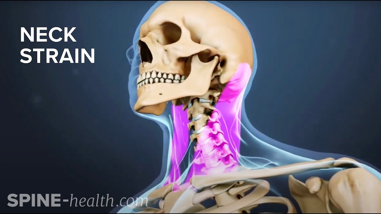
Neck Strain Causes And Remedies from i.ytimg.com Learn more about head and neck anatomy, including the top part of the skeleton, muscles, and more with our digital flashcards. Click to view large image. The pll starts at c2 and goes down the back of the vertebral bodies and intervertebral discs. A collection of anatomy notes covering the key anatomy concepts that medical students need to learn. Want to learn more about it? Dummies helps everyone be more knowledgeable and confident in applying what they know. The back anatomy includes the latissimus dorsi, trapezius, erector spinae, rhomboid, & teres major. Muscles of the posterior neck and the back.
Learn about these muscles, their locations & functional the traps are quite a complex set of muscles.
Neck muscles help support the cervical spine and contribute to movements of the head, neck, upper back, and posterior longitudinal ligament (pll). This article concerning the anatomy of the head and neck area gives you a clear structure at hand to see anatomy and function of the regions of the lower face. A dynamic and interactive atlas of ent imaging. Dummies helps everyone be more knowledgeable and confident in applying what they know. This mri neck axial cross sectional anatomy tool is absolutely free to use. A collection of anatomy notes covering the key anatomy concepts that medical students need to learn. The longus capitis and rectus capitis anterior are the direct antagonists of the muscles at the back of the neck, serving to restore the head to its natural position after it has been drawn backward. Anatomical observation and palpation sk… surface anatomy: The majority of these nerves control the functions of the upper extremities and allow you to feel your arms, shoulder, and back of your head. From the sides and the back of the neck, the splenius capitis inserts onto the head region, and the splenius. Surface anatomy and surface markings bibliographic record list of illustrations subject index. The anterior jugular vein (v. Cervical spine anatomy is quite complex.
The majority of these nerves control the functions of the upper extremities and allow you to feel your arms, shoulder, and back of your head. This entry was posted in anatomy by admin. The longus capitis and rectus capitis anterior are the direct antagonists of the muscles at the back of the neck, serving to restore the head to its natural position after it has been drawn backward. It runs down the back part of the neck, and opens into the external jugular vein just below the middle of its course. This mri neck axial cross sectional anatomy tool is absolutely free to use.
Spine Structure Function Parts Segments Spine Problems Spine Health from www.clevelandclinic.org It runs down the back part of the neck, and opens into the external jugular vein just below the middle of its course. When most people mention their back, what they are actually referring to is their spine. It is important to know what normal is so you can recognize… anatomical observation and palpation skills are necessary for… The head rests on the top part of the vertebral column, with the skull joining at c1. Whether it's to pass that big test, qualify for that big promotion or even master that cooking technique; Clinically, surface anatomy is used to split the neck into anterior and posterior triangles which provide clues as to the location of specific structures. The splenius muscles originate at the midline and run laterally and superiorly to their insertions. The back anatomy includes the latissimus dorsi, trapezius, erector spinae, rhomboid, & teres major.
« back show on map ».
This mri neck axial cross sectional anatomy tool is absolutely free to use. 3d interactive tutorials on the anatomy of the neck, including the anatomical organisation, musculature, larynx, pharynx, blood supply and innervation. The longus capitis and rectus capitis anterior are the direct antagonists of the muscles at the back of the neck, serving to restore the head to its natural position after it has been drawn backward. Use the mouse scroll wheel to move the images up and down alternatively use the tiny arrows (>>) on both side of the image to move the images. It is made up of bones, discs, muscles, ligaments, nerves and tendons. Click to view large image. This entry was posted in anatomy by admin. The pll starts at c2 and goes down the back of the vertebral bodies and intervertebral discs. The splenius muscles originate at the midline and run laterally and superiorly to their insertions. The neck is the area between the skull base and the clavicles. Anatomy of the head and neck: Surface anatomy and surface markings bibliographic record list of illustrations subject index. Some important structures contained in or passing through the neck include the seven cervical vertebrae and enclosed spinal cord, the jugular veins and carotid arteries, part of the esophagus, the larynx.
Learn about these muscles, their locations & functional the traps are quite a complex set of muscles. It runs down the back part of the neck, and opens into the external jugular vein just below the middle of its course. This article concerning the anatomy of the head and neck area gives you a clear structure at hand to see anatomy and function of the regions of the lower face. The neck is the area between the skull base and the clavicles. All of the anatomical structures of the face with labels on 150 axial and coronal slices from a scan:
Occipital Neuralgia Causes Symptoms Diagnosis And Treatment from www.aans.org The splenius muscles originate at the midline and run laterally and superiorly to their insertions. The neck is the area between the skull base and the clavicles. The longus capitis and rectus capitis anterior are the direct antagonists of the muscles at the back of the neck, serving to restore the head to its natural position after it has been drawn backward. The pll starts at c2 and goes down the back of the vertebral bodies and intervertebral discs. This article describes the anatomy of the head and neck of the human body, including the brain, bones, muscles, blood vessels, nerves, glands, nose, mouth, teeth, tongue, and throat. A dynamic and interactive atlas of ent imaging. Anatomists tend to classify the body into during muscle traction, the cheeks are pulled together, which makes food move back and forth. Click to view large image.
Use the mouse scroll wheel to move the images up and down alternatively use the tiny arrows (>>) on both side of the image to move the images.
How to view the anatomical labels. This article looks at the anatomy of the back, including bones, muscles, and nerves. Understanding the anatomy of your cervical spine and the vital nerves it contains should motivate you to adopt behaviors that help prevent neck injury and. Bones of the neck picture. This article concerning the anatomy of the head and neck area gives you a clear structure at hand to see anatomy and function of the regions of the lower face. The pll starts at c2 and goes down the back of the vertebral bodies and intervertebral discs. From the sides and the back of the neck, the splenius capitis inserts onto the head region, and the splenius. 17.11.2015 · neck anatomy explained the neck begins at the base of the skull and connects to the thoracic spine (the upper back). An overview of the anatomy of the hand, including the bones of the hand, muscles, blood supply and nerve supply. Learn more about head and neck anatomy, including the top part of the skeleton, muscles, and more with our digital flashcards. The anterior jugular vein (v. Click now to study the muscles, glands and organs of the neck at kenhub! Dummies helps everyone be more knowledgeable and confident in applying what they know.


0 Comments