Anatomical Name Of Lower Back Muscles - Back Strains And Sprains : The superficial back muscles are covered by skin.. Which are linked to a breakdown of each muscle with anatomical position: The latter group is the intrinsic muscle group. Your lower back (lumbar spine) is the anatomic region between your lowest rib and the upper part of the buttock.1 your spine in this region. Name the 4 muscles of the quadriceps femoris group. The superficial back muscles are the muscles found just under the skin.
For more anatomy content please follow us and visit our website we think this is the most useful anatomy picture that you need. Related posts of muscle names of lower back. This article will focus on the superficial group. In this position, the body is straight in standing position with eyes also looking straight. Chest muscle anatomy exercises 12 photos of the chest muscle anatomy exercises chest muscle anatomy and exercises, chest muscle anatomy exercises, human muscles, chest muscle anatomy.
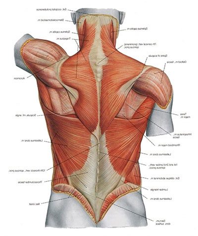
The 5 Types Of Back Pain Your Guide To Identifying Your Back Condition from cornerstonephysio.com This article covers the anatomy of the superficial muscles of the back, including trapezius, latissimus dorsi, levator scapulae, rhomboid major and minor. This article will focus on the superficial group. The back muscles stabilize and move the vertebral column, and are grouped according to the lengths similar to the erector spinae muscles, the semispinalis muscles in this group are named for the. The muscles of the back can be divided in three main groups according to their anatomical position and function. Back / anatomy & histology*. Which are linked to a breakdown of each muscle with anatomical position: We hope this picture muscles of lower back diagram can help you study and research. Name the 4 muscles of the quadriceps femoris group.
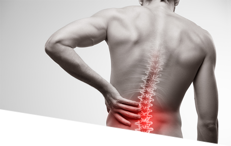
Your lower back (lumbar spine) is the anatomic region between your lowest rib and the upper part of the buttock.1 your spine in this region.
Your lower back (lumbar spine) is the anatomic region between your lowest rib and the upper part of the buttock.1 your spine in this region. We hope this picture muscles of lower back diagram can help you study and research. The subcostal muscles are strips of muscle located on the internal surface of the lower ribs, sharing a plane it separates the thoracic and abdominal cavities and facilitates the passage of anatomical for descriptive purposes, the muscles of the back are divided into two groups; This article will focus on the superficial group. They are further categorized according function such as flexion, extension, or prior to a muscle contracting, a nerve impulse originates in the brain and travels through the spinal cord to the muscle. Back / anatomy & histology*. This is the position in which the back of the body is directed upwards. The latter group is the intrinsic muscle group. The muscles of the back that work together to support the spine, help keep the body upright and allow twist and bend in many directions. This is a compound exercise that also involves the triceps and the front deltoids, also recruits the upper and lower back muscles, and traps. There are many muscles that help to both move and stabilize the spine. Name the 4 muscles of the quadriceps femoris group. Chest muscle anatomy exercises 12 photos of the chest muscle anatomy exercises chest muscle anatomy and exercises, chest muscle anatomy exercises, human muscles, chest muscle anatomy.
To simplify things, i'm going to split the back into three sections; In this position, the body is straight in standing position with eyes also looking straight. In anatomical terminology, chewing is called mastication. These muscles, including the gluteus maximus and the hamstrings, extend the thigh at the hip in support of the body's weight and propulsion. Each of these fibres play a specific role, giving the trapezius muscle many roles.
Muscles Of The Lower Back Anatomy Anatomy Drawing Diagram from www.spinalbackrack.com Muscles that move the leg are located in the thigh region. Neck muscle anatomy ultrasound 12 photos of the neck muscle anatomy ultrasound , human muscles. Learn anatomical details of the lower back muscles, so you can draw them. In this position, the body is straight in standing position with eyes also looking straight. Structural groups of muscles largely determine functional groups—that is, the structural location of a muscle largely determines its mover function. Muscles are described using unique anatomical terminology according to their actions and structure. These muscles, including the gluteus maximus and the hamstrings, extend the thigh at the hip in support of the body's weight and propulsion. The muscles located in the leg that move the ankle and foot are divided into anterior, posterior, and lateral compartments.
Neck muscle anatomy ultrasound 12 photos of the neck muscle anatomy ultrasound , human muscles.
Energy is needed for the. Structural groups of muscles largely determine functional groups—that is, the structural location of a muscle largely determines its mover function. The superficial back muscles are the muscles found just under the skin. Below we have a list of muscle names. Understanding the anatomy of your lower spine can help you communicate more effectively with the medical professionals who treat your lower back pain. Learn how to draw the lower back muscles by learning their form. The muscles of the back can be divided in three main groups according to their anatomical position and function. A discussion of the lower back wouldn't be complete without an overview of the sacrum. Name the 4 muscles of the quadriceps femoris group. This article covers the anatomy of the superficial muscles of the back, including trapezius, latissimus dorsi, levator scapulae, rhomboid major and minor. There are many muscles that help to both move and stabilize the spine. Last time we learned the anatomical details of the lower back muscles. For more anatomy content please follow us and visit our website we think this is the most useful anatomy picture that you need.
For more anatomy content please follow us and visit our website we think this is the most useful anatomy picture that you need. The muscles of the back that work together to support the spine, help keep the body upright and allow twist and bend in many directions. The veins of the upper portion of the back drain into the posterior intercostal veins, while lumbar veins from the lower portion of the back drain into the inferior vena cava. Related posts of muscle names of lower back. Understanding the anatomy of your lower spine can help you communicate more effectively with the medical professionals who treat your lower back pain.
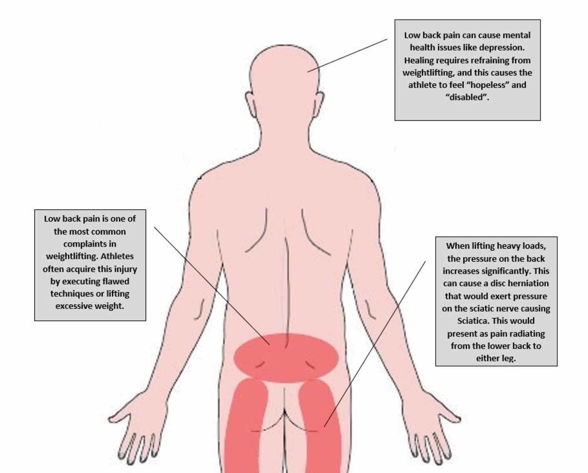
Cureus Low Back Pain Among Weightlifting Adolescents And Young Adults from assets.cureus.com The anatomy of the lumbar spine is quite complex. Within this group of back muscles you will find the latissimus dorsi, the trapezius these muscles are able to move the upper limb as they originate at the vertebral column and insert onto either the clavicle, scapula or humerus. This is the position in which the back of the body is directed upwards. Muscles are described using unique anatomical terminology according to their actions and structure. Anatomy muscles of lower body. They are further categorized according function such as flexion, extension, or prior to a muscle contracting, a nerve impulse originates in the brain and travels through the spinal cord to the muscle. Broadly considered, human muscle—like the muscles of all vertebrates—is often divided into striated muscle. The latter group is the intrinsic muscle group.
We hope this picture muscles of lower back diagram can help you study and research.
Below we have a list of muscle names. Your lower back (lumbar spine) is the anatomic region between your lowest rib and the upper part of the buttock.1 your spine in this region. To simplify things, i'm going to split the back into three sections; This lesson covers the erector spinae and latissimus dorsi muscles. Here the extrinsic back muscles are classified into logical subgroups to facilitate knowledge. Each of these fibres play a specific role, giving the trapezius muscle many roles. Muscles are described using unique anatomical terminology according to their actions and structure. The muscles of the back can be divided in three main groups according to their anatomical position and function. Neck muscle anatomy ultrasound 12 photos of the neck muscle anatomy ultrasound , human muscles. You can click the image to magnify if you cannot see clearly. Muscles are named according to their shape, location, or a combination. Chest muscle anatomy exercises 12 photos of the chest muscle anatomy exercises chest muscle anatomy and exercises, chest muscle anatomy exercises, human muscles, chest muscle anatomy. In anatomical terminology, chewing is called mastication.


0 Comments